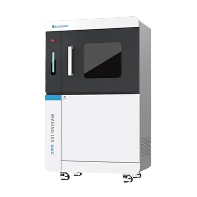
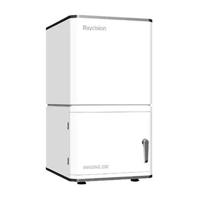
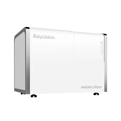
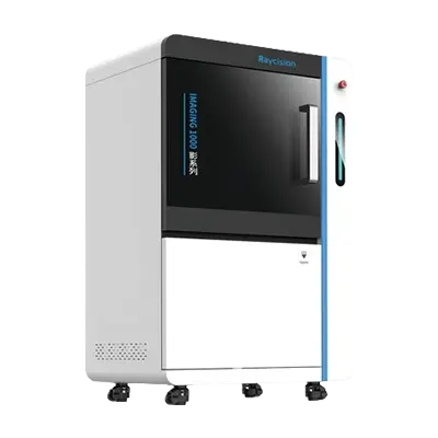
Raycision offers state-of-the-art preclinical in vivo imaging systems designed to enable researchers to visualize and monitor biological processes in live animals, particularly small mammals such as mice and rats, in a non-invasive and highly detailed manner. These advanced imaging systems integrate multiple modalities, including bioluminescence, fluorescence, and CT, providing comprehensive insights into anatomical, functional, and molecular dynamics within the living organism. By facilitating real-time, longitudinal studies, Raycision’s technology allows for the continuous observation of disease progression, drug distribution, and therapeutic efficacy without the need for euthanizing animals at various stages, enhancing the accuracy and reliability of experimental data while aligns with ethical standards in animal research.


High-Resolution Imaging:
Micro-CT provides high-resolution, three-dimensional images of small animals, allowing for detailed visualization of anatomical structures. This is particularly useful for studying bone morphology, lung architecture, and other dense tissues with exceptional clarity.
Non-Invasive and Longitudinal Studies:
Micro-CT imaging is non-invasive, enabling researchers to perform longitudinal studies on the same animal over time. This reduces the number of animals required for experiments and allows for the monitoring of disease progression, treatment effects, and recovery processes in real-time.
3D Reconstruction:
The ability to reconstruct three-dimensional images from micro-CT data provides a comprehensive view of the internal structures of small animals. This 3D visualization is invaluable for understanding complex anatomical relationships and spatial distributions of tissues and organs.
Quantitative Analysis:
The high-resolution images obtained from micro-CT can be quantitatively analyzed to measure changes in tissue density, volume, and structure. This quantitative data is crucial for assessing the efficacy of treatments, understanding disease mechanisms, and conducting detailed phenotypic studies.
Integration with Other Modalities:
Micro-CT can be integrated with other imaging modalities, such as optical molecular imaging, to provide complementary information. This multimodal approach enhances the overall understanding of biological processes by combining anatomical, functional, and molecular data.
Multimodality imaging in animal models of diseases offers a range of significant advantages that enhance the depth, accuracy, and comprehensiveness of biomedical research. By combining different imaging techniques, researchers can obtain a more holistic view of biological processes and disease mechanisms.
Comprehensive Data Acquisition:
Multimodality imaging allows for the simultaneous acquisition of anatomical, functional, and molecular data. For example, combining CT (which provides high-resolution anatomical details) with optical molecular imaging (which offers functional and metabolic information) enables a more complete understanding of disease states and therapeutic effects.
Enhanced Sensitivity and Specificity:
Different imaging modalities have unique strengths and limitations. By integrating multiple modalities, researchers can leverage the strengths of each technique to improve overall sensitivity and specificity. This leads to more accurate detection and characterization of disease processes.
Reduced Animal Use:
By obtaining comprehensive data from a single animal using multiple imaging modalities, researchers can reduce the number of animals needed for experiments. This aligns with ethical guidelines and the principles of the 3Rs (Replacement, Reduction, and Refinement) in animal research.
Cross-Validation of Data:
Multimodality imaging enables cross-validation of data obtained from different techniques. This helps confirm findings and reduces the likelihood of artifacts or errors, leading to more robust and reliable results.
Accelerated Drug Development:
In drug development, multimodality imaging can accelerate the evaluation of new therapies by providing comprehensive data on drug distribution, target engagement, and therapeutic efficacy. This can lead to more informed decision-making and faster progression through the preclinical pipeline.
In summary, multimodality imaging in animal models of diseases offers a powerful approach to obtaining comprehensive, accurate, and detailed insights into biological processes and disease mechanisms. By combining the strengths of different imaging techniques, researchers can enhance the quality of their data, reduce animal use, and accelerate the development of new therapies.
Tel:
E-mail:
Address:
Zhong'an Chuanggu Technology Park, Hefei, China