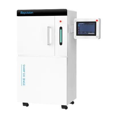
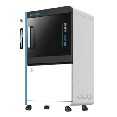
Raycision's preclinical image-guided X-ray radiation systems are designed to deliver precise radiation therapy to small animal models, such as mice and rats. These systems provide high-resolution images, image-guided radiation delivery, adjustable radiation dose, 3d treatment planning, and treatment response evaluation, making them ideal tools for studying the effects of radiation on biological tissues, develop new cancer treatments, and understand disease mechanisms.


No radioactive material: X-ray irradiators do not use radioactive isotopes, such as Cobalt-60 or Cesium-137, which are used in gamma ray irradiators. This eliminates the risks associated with handling, storing, and disposing of radioactive materials.
Reduced regulatory burden: the absence of radioactive materials simplifies regulatory compliance, reduces environmental impact, and mitigates security risks related to the theft or misuse of radioactive substances.
On-demand operation: The X ray irradiator system can be turned on and off as needed, providing immediate availability and reducing the risk of accidental exposure.
Adjustable dose rates: X-ray irradiators allow for precise control over dose rates and energy levels, enabling tailored irradiation protocols. They also have lower maintenance costs, as they do not require periodic replacement of decaying radioactive sources.
The dose calculation algorithm in preclinical image guided radiation systems is a critical component that ensures precise and accurate delivery of radiation to the target area while minimizing exposure to surrounding healthy tissues. These algorithms integrate imaging data with radiation delivery parameters to create a detailed treatment plan. The following are some key components of dose calculation algorithms.
Acquisition of high-resolution images: high-resolution images provide detailed anatomical and, in some cases, functional information about the target area and surrounding tissues.
Target delineation: the target area (e.g., tumor) and critical structures (e.g., organs at risk) are delineated or contoured on the imaging data.
Volume definition: the delineated structures are used to define the volumes that will receive specific doses of radiation.
Beam modeling: the characteristics of the radiation beam, such as energy, intensity, and shape, are modeled.
Treatment planning: The calculated dose distribution of the small animal irradiator is used to create a treatment plan that optimizes the delivery of radiation to the target while minimizing exposure to healthy tissues.
Tel:
E-mail:
Address:
Zhong'an Chuanggu Technology Park, Hefei, China